Lung cancer resection surgeries and their post operative appearance
Surgical resection is the mainstay of treatment for fit and willing patients with early NSCLC.
The type and extent of resection is dependent on the T-stage of the tumour,
its location and the patient’s pulmonary function(2).
The goal is an adequate resection while preserving as much lung function as possible.
The TNM staging system for NSCLC is displayed in Table 1.

Table 1: 8th edition of the AJCC TNM staging system for Non–Small Cell Lung Cancer
Lobectomy
Lobectomy was established as the gold standard for early stage (T1-2,N0) tumours not invading the main airway in 1995(3).
Lobectomy involves removal of the abnormal lobe and its surrounding pleura following transection of the lobar bronchus and artery(1)(Figure 1).
The bronchial stump is covered with viable tissue,
such as a pleural flap,
to prevent dehiscence.
VATS (video assisted thorascopic surgery) lobectomy is a less invasive approach,
associated with lower post-operative pain,
perioperative complications and shorter hospital stay. Disease free and overall survival rates are comparable for open and VATS lobectomy(4).

Fig. 1: Illustration of a right upper lobectomy.
Post-operative chest radiographs show volume loss and a chest drain,
typically removed on day 3/4.
Ipsilateral lung changes are common.
Development of contralateral lung changes should raise suspicion of clinical deterioration.
The volume loss matures over one year and stabilises (Figure 2).

Fig. 2: Post lobectomy chest radiograph appearances. (A) Pre-op. Chest radiograph demonstrates a right lower lobe adenocarcinoma (arrow). (B) Day 1 post-op. AP portable chest radiograph with a right wide bore chest drain in situ and subcutaneous emphysema overlying the right chest wall. (C) Day 3 post-op. The chest drain has been removed and there are minor ipsilateral parenchymal changes. Contralateral lung remains clear. (D) One month post-op. Right volume loss. (E) One year post-op. The right volume loss is established and stable. Unfortunately this patient developed a rib metastasis (circle).
CT permits direct visualisation of the bronchial stump (Figure 3).
Volume loss is evident with elevation of the hemidiaphragm or mediastinal shift.
Post-operative pleural thickening is common and is thicker if there has been chest wall resection.

Fig. 3: Post-operative CT thorax. (A) CT Thorax demonstrates the bronchial stump/suture line. (B) Left upper pleural thickening and volume loss.
Sublobar resection
Sublobar resection involves a segmentectomy: removal of the tumour and associated lung segments,
the segmental bronchus and artery (Figure 4).
While it was previously thought that sublobar resections should be reserved for those unfit for lobectomy,
there is emerging evidence that patients with non-invasive/minimally invasive tumours would benefit.
Comparable outcomes from lobectomies and sublobar resections are possible in a select cohort of patients(5).
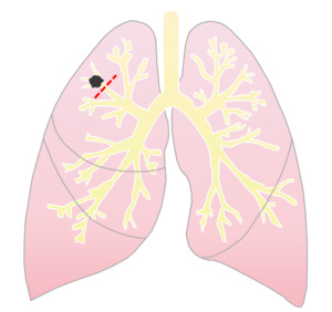
Fig. 4: Illustration of a sublobar resection/segmentectomy
CT appearance is similar to post-lobectomy except the volume loss is less pronounced and there is a suture line extending back to the hilum (Figure 5).

Fig. 5: (A + B) CT Thorax post right apical lower lobe segmentectomy. The suture line extends from the periphery to the hilum.
Sleeve lobectomy
In cases where tumour has spread to the bronchial tree,
sleeve lobectomy can be performed to avoid pneumonectomy(6).
It is most commonly used for right upper lobe resections due to the long length of the bronchus intermedius,
which permits anastomosis.
The diseased lobe is resected,
along with a portion of the common airway,
followed by end-to-end anastomosis of the distal and proximal airway(1). The morbidity and mortality of a sleeve resection is similar to that for pneumonectomy but better functional lung remains(7).
Chest radiograph following sleeve lobectomy is similar to post-lobectomy.
CT shows the bronchial anastomosis.
Pneumonectomy
Pneumonectomy is performed for multifocal,
bulky or central disease(2).
The classical technique,
intrapleural pneumonectomy,
involves transection of the main bronchus and pulmonary artery/veins,
removal of the entire lung and its visceral pleura (Figure 6).
The bronchial stump is reinforced with vascularised tissue to prevent dehiscence.

Fig. 6: Illustration of a right pneumonectomy.
Post-operative radiographs show the empty post-pneumonectomy space on day 1.
Over the following days the space gradually fills with fluid with no drop in the level.
By day 7 the space should be approximately 70-80% full.
The fluid level is close to the top of the hemithorax by patient discharge,
typically at 10-14 days.
Once the space fills we see progressive volume loss on the pneumonectomy side (Figure 7).

Fig. 7: Post Pneumonectomy X-ray changes. (A) CT Thorax demonstrates central left hilar tumour for which pneumonectomy is indicated. (B) Day 1 post-op. Chest radiograph shows empty left hemithorax. (C-E) Gradual filling of the left post pneumonectomy space during the early post-operative period. (F) One year post-op. Opacified left hemithorax with volume loss.
The degree of volume loss post pneumonectomy is variable (Figure 8).

Fig. 8: (A) Right pneumonectomy with right-sided mediastinal shift and tracheal deviation. (B) Left pneumonectomy with a centrally positioned trachea.
On CT,
the pneumonectomy space should fill with fluid (Figure 9).
Pockets of gas can be seen within the space in the first few weeks but significant air indicates a bronchopleural fistula.
There is pleural thickening and enhancement within the space and therefore assessment for infection can be challenging.

Fig. 9: CT Thorax following left pneumonectomy demonstrates fluid within the post pneumonectomy space with volume loss and left mediastinal shift. There is pleural enhancement and thickening (arrows).
Extrapleural pneumonectomy is for locally advanced or pleural based tumours.
It includes resection of the lung,
its visceral/parietal/mediastinal pleura,
and the associated pericardium and diaphragm.
Extrapleural approach may be identified on post-operative imaging by the presence of a synthetic pericardial or diaphragmatic graft or vascular stents(1)(Figure 10).

Fig. 10: CT appearance post extrapleural pneumonectomy. (A) The pericardium is reinforced with a synthetic graft. B) Extensive mediastinal resection requires insertion of a vascular stent.
Complications
Mortality rates post limited lung resections are typically low however increase for patients undergoing lobectomy,
ranging from 1-4%(8),
and can be as high as 11% post pneumonectomy(1).
Post-operative morbidity rates also depend on the extent of resection,
as well as the patients' pre-morbid status,
and vary from 20-60%(9).
As well as general post-operative complications including haemorrhage,
infection,
and pulmonary embolism,
there are specific complications associated with lung resection that can be divided into early post-operative complications,
in the initial post-operative period,
and late post-operative complications,
post discharge (Table 2).

Table 2: Specific post lung resection complications
Early Complications
Acute Lung Injury (ALI)/ Acute Respiratory Distress Syndrome (ARDS)
ALI/ARDS occurs in 2-5% patients and mortality can be as high as 80%(10).
Median time of onset is 4 days post-operatively(11).
Imaging features of ALI and ARDS are indistinguishable and while the appearances are characteristic,
they are not specific with pulmonary oedema having a very similar appearance(1).
Findings include diffuse bilateral groundglass opacities and development of consolidation which demonstrates an anteroposterior density gradient,
with more dense consolidation in the dependent lung (Figure 11).
The absence of a pleural effusion is more consistent with ARDS than pulmonary oedema.

Fig. 11: Acute Lung Injury. (A) Day 1 post left lobectomy with left chest drain in situ and minor left parenchymal changes. B) Day 3 post-op. Developing right lower zone parenchymal changes. C) Day 5 post-op. Worsening of the contralateral right lung changes. D) CT Thorax demonstrates extensive bilateral groundglass opacities with more confluent consolidation dependently. There is no pleural effusion. Findings are consistent with an acute lung injury/ARDS.
Post Resection Pulmonary Oedema
Pulmonary oedema occurs more commonly in patients post pneumonectomy than post lobectomy and affects approximately 2-5% of patients(12).
It more commonly occurs following a right pneumonectomy(13).
It can be clinically and radiologically similar to ALI and can occur at any time post-operatively however increasing time from surgery makes pulmonary oedema more likely as ALI tends to occur in the first 3-4 days.
The presence of an effusion can also help distinguish oedema from ALI.
Mortality is high and can be prevented by avoiding excessive fluid replacement and transfusions(12).

Fig. 12: Post resection pulmonary oedema. (A) Day 1 post pneumonectomy shows empty space in the left hemithorax and a clear right lung. (B) Day 7 post-op. Increasing fluid level in the left hemithorax. New right lower zone opacification. (C) Day 9 post-op. Diffuse right lung airspace opacification with increased interstitial markings and a right pleural effusion. Although it looks similar to an ALI, the timing is too late and this represents pulmonary oedema.
Stump Dehiscence/ Bronchopleural Fistula (BPF)
Bronchial stump dehiscence refers to the breakdown of the bronchial stump/suture and subsequent BPF.
It occurs in approximately 5% of pneumonectomy patients,
more commonly than post lobectomy.
Mortality,
typically due to aspiration and development of ARDS,
occurs in up to 25%(1).
They can occur early or late and are more common on the right(12).
If early,
they tend to occur in the first week and usually relate to poor surgical technique.
These tend to be large and fail to respond to conservative management.
Early BPF can present with increased output from the chest drain,
accumulation of air within the pleural space (Figure 13) or failure of a pneumonectomy space to fill with fluid/ drop of >2cm in the fluid level (Figure 14).

Fig. 13: Bronchopleural fistula. (A) Day 2 post right sleeve lobectomy. The patient is recovering well. The chest drain showed little output and was removed on day 3. (B) Day 4 post-op. Patient became unwell and chest x-ray demonstrates a large collection of air in the pleural space (arrows). (C) Day 5 post-op. Chest drains were reinserted and there was a very large output immediately. The pleural accumulation of air persisted. Findings were consistent with a bronchial stump dehiscence/bronchopleural fistula.

Fig. 14: Bronchial stump Dehiscence/ BPF post pneumonectomy. (A-C) Patient progressing well with increasing fluid level in the left hemithorax post left completion pneumonectomy (previous left lower lobectomy for squamous cell carcinoma). (D) On Day 5 the fluid level suddenly drops and continues to do so on Day 7 (E). (F) Pigtail catheter inserted on Day 8. Findings are consistent with bronchial stump dehiscence.
Most patients will not respond to chest drain insertion.
Surgical intervention is typically required,
involving the refashioning and reinforcement of the bronchial stump with a vascularised tissue flap like omentum or muscle.
Surgical intervention is successful in almost 90% of patients(1).
Persistent air leaks
The main differential diagnosis for a BPF is prolonged air leaks.
Up to 50% of patients will have some form of leak however many resolve within hours/days.
A persistent air leak is one that is present after 4-5 days(16).
These have a relatively small amount of pleural air in comparison to BPF (Figure 15).
Predisposing factors include upper lobectomy,
damage to an area of lung intra-operatively and incomplete fissures which are incompletely stapled(16).
Management with chest tube drainage is typically successful.
Occasionally pleurodesis or surgical intervention may be required.

Fig. 15: Persistent air leak. (A) Day 1 post right upper lobectomy. Right chest drain in situ with an apical right pneumothorax. (B) Day 3 post-op. Right pneumothorax slightly increases in size. (C) Day 4 post-op. Persistent right apical pneumothorax consistent with a persistent air leak.
Empyema
Empyema is a potentially fatal complication and is reported to occur in 1-10% of patients following lung resection (9).
They can occur early or late and are associated with BPF in approximately 80% of cases (1).
In the early phase,
if not secondary to BPF,
it is related to contamination of the surgical field.
Empyema presents with signs of sepsis,
pyrexia,
elevated inflammatory markers and a deterioration in the patient’s clinical state (1).
Chest radiograph may demonstrate a simple or loculated pleural effusion.
CT shows fluid within the post surgical space,
which is typically complex,
associated with pleural thickening and enhancement and may exert mass effect on the mediastinum (Figre 16).

Fig. 16: Empyema. (A) Day 9 post right lower lobectomy. Chest radiograph demonstrates a loculated pleural effusion. (B) CT Thorax reveals an air pocket (star) with a surrounding loculated effusion. There is pleural enhancement (arrow). Findings are consistent with an empyema.
Initial treatment involves chest tube drainage with intravenous antibiotics.
Complete sterilisation can be very difficult.
An eloesser flap is a single stage procedure for the treatment of severe pleural empyema,
and involves a U-shaped incision and the resection of a number of adjacent posterolateral ribs.
The U-shaped flap is then folded into the pleural space creating a permanent communication and allowing one way drainage of the empyema (Figure 17) (1).

Fig. 17: Coronal and axial CT images of an Eloesser Flap.
Chylothorax
Chylothorax occurs in approximately 1% of patients post pneumonectomy and is more common than in lobectomy patients(12).
Injury to the thoracic duct or one of the main lymphatic ducts results in accumulation of chyle in the pleural space.
Chest radiograph may reveal a rapidly increasing fluid level in the left post pneumonectomy space causing mediastinal shift to the right (Figure 18).
Chest drain insertion with sampling of the fluid is required for diagnosis with the appearance of the fluid and triglyceride content confirming the diagnosis(8).

Fig. 18: Chylothorax. (A) Day 4 post left pneumonectomy. The pneumonectomy space is filling with fluid as expected. (B) Day 6 post-op. The left pneumonectomy space has rapidly filled with fluid and is associated with mass effect and mediastinal shift to the right. )C) CT Thorax shows the mass effect and mediastinal shift. Percutaneous sampling confirmed the presence of a chylothorax.
Lobar torsion.
Lobar torsion is a rare early post-operative complication following lobectomy,
occurring in less than 0.2% of patients(17).
The middle lobe is most commonly affected as it moves cephalad after a right upper lobectomy.
There is little resistance to movement of the remaining lung and as the lobes move to fill the space,
the affected lobe twists on its vascular pedicle at the hilum.
This causes occlusion of the bronchus and pulmonary arteries resulting in venous obstruction and haemorrhagic infarction.
Typically the patient’s clinical course deteriorates significantly.
They become hypoxic.
Raised inflammatory markers and pyrexia may also be present.
Chest radiograph demonstrates increasing density and can mimic atelectasis/collapse.
CT shows the torted oedematous lobe and occlusion of the bronchus and artery.
Urgent surgical exploration is required (Figure 19)(1).

Fig. 19: Middle lobe torsion. (A) Day 2 post right upper lobectomy. Patient complained of chest pain but CTPA was negative. (B) Day 9 post-op. Chest radiograph revealed a well defined opacity in the right upper zone with volume loss suggestive of lobar collapse. (C) CT reveals an oedematous expanded middle lobe.
References: Royal Brompton Hospital
Late complications
Delayed BPF and empyema.
Late BPF occur secondary to recurrent infections or malignancy and are also associated with empyema.
If the bronchial stump remains intact,
empyema is due to haematogenous spread of infection.
Radiological appearances are similar to that of early BPF with the appearance of new air in the pneumonectomy space (Figure 20).
Management of late BPF with empyema,
particularly in ventilated patients,
is difficult.
Some will respond to chest drain insertion however a tissue flap inserted to close the fistula and sterilise the space is sometimes required.

Fig. 20: Delayed BPF and empyema. 60 year old man 3 years post pneumonectomy for squamous cell carcinoma with recurrent chest infections. CT Thorax performed during acute deterioration revealed new air in the pneumonectomy space.
Oesophagopleural Fistula
Oesophagopleural fistula is due to recurrent tumour or chronic inflammation and typically occurs within the first two years post surgery(9).
Chest radiograph demonstrates findings similar to that of BPF however CT Thorax allows visualisation of the actual fistula.
Leak of oral contrast into the pleural space during an upper GI contrast swallow also demonstrates the fistula (Figure 21).

Fig. 21: Oesophagopleural Fistula. (A) 2 years post lobectomy. Patient presented with sepsis and recurrent chest infections. CT revealed new air in the pleural space. (B) A water soluble contrast swallow revealed leak of contrast from the oesophagus into the right pleural space consistent with oesophagopleural fistula.
Post pneumonectomy syndrome
Post pneumonectomy syndrome is an uncommon complication that typically occurs within one year post-op and is more common in children and young adults.
It is more common following a right pneumonectomy and presents with shortness of breath and recurrent infections.
After a right pneumonectomy,
the heart and mediastinum move posterolaterally into the pneumonectomy space.
The distal trachea and left main bronchus rotate to the right and the left main bronchus is compressed between the left main pulmonary artery and the aorta (Figure 22)(12).

Fig. 22: Post pneumonectomy syndrome. 68 year old man presents with increasing shortness of breath post pneumonectomy 4 years previously. (A) CXR shows volume loss with tracheal deviation. (B+C) CT demonstrates significant mediastinal shift with narrowing of the left main bronchus by the left pulmonary artery consistent with post pneumonectomy syndrome.
Recurrence
There is no internationally accepted protocol for CT surveillance following lung cancer resection and follow-up is typically performed as per individual department protocols.
In order to assess for recurrence one most know the expected post-operative appearance.
Recurrence may be local (26%),
distant (44%) or a combination of both (30%)(18).
New nodal disease,
recurrence at the bronchial stump,
pulmonary nodules,
either ipsilateral or contralateral,
and pleural recurrence,
are typical findings of intrathoracic recurrence (Figures 23/24).

Fig. 23: Patterns of recurrence. (A) New right paratracheal lymph node. (B) New ipsilateral pulmonary nodule. (C) Pleural recurrence with rib destruction.

Fig. 24: Bronchial stump recurrence. CT (A – C) shows gradually increasing soft tissue at the bronchial stump over serial yearly CTs.


 Pre-op. Chest radiograph demonstrates a right lower lobe adenocarcinoma (arrow). (B) Day 1 post-op. AP portable chest radiograph with a right wide bore chest drain in situ and subcutaneous emphysema overlying the right chest wall. (C) Day 3 post-op. The chest drain has been removed and there are minor ipsilateral parenchymal changes. Contralateral lung remains clear. (D) One month post-op. Right volume loss. (E) One year post-op. The right volume loss is established and stable. Unfortunately this patient developed a rib metastasis (circle).")
 CT Thorax demonstrates the bronchial stump/suture line. (B) Left upper pleural thickening and volume loss.")

 CT Thorax post right apical lower lobe segmentectomy. The suture line extends from the periphery to the hilum.")

 CT Thorax demonstrates central left hilar tumour for which pneumonectomy is indicated. (B) Day 1 post-op. Chest radiograph shows empty left hemithorax. (C-E) Gradual filling of the left post pneumonectomy space during the early post-operative period. (F) One year post-op. Opacified left hemithorax with volume loss.")
 Right pneumonectomy with right-sided mediastinal shift and tracheal deviation. (B) Left pneumonectomy with a centrally positioned trachea.")
.")
 The pericardium is reinforced with a synthetic graft. B) Extensive mediastinal resection requires insertion of a vascular stent.")

 Day 1 post left lobectomy with left chest drain in situ and minor left parenchymal changes. B) Day 3 post-op. Developing right lower zone parenchymal changes. C) Day 5 post-op. Worsening of the contralateral right lung changes. D) CT Thorax demonstrates extensive bilateral groundglass opacities with more confluent consolidation dependently. There is no pleural effusion. Findings are consistent with an acute lung injury/ARDS.")
 Day 1 post pneumonectomy shows empty space in the left hemithorax and a clear right lung. (B) Day 7 post-op. Increasing fluid level in the left hemithorax. New right lower zone opacification. (C) Day 9 post-op. Diffuse right lung airspace opacification with increased interstitial markings and a right pleural effusion. Although it looks similar to an ALI, the timing is too late and this represents pulmonary oedema.")
 Day 2 post right sleeve lobectomy. The patient is recovering well. The chest drain showed little output and was removed on day 3. (B) Day 4 post-op. Patient became unwell and chest x-ray demonstrates a large collection of air in the pleural space (arrows). (C) Day 5 post-op. Chest drains were reinserted and there was a very large output immediately. The pleural accumulation of air persisted. Findings were consistent with a bronchial stump dehiscence/bronchopleural fistula.")
 Patient progressing well with increasing fluid level in the left hemithorax post left completion pneumonectomy (previous left lower lobectomy for squamous cell carcinoma). (D) On Day 5 the fluid level suddenly drops and continues to do so on Day 7 (E). (F) Pigtail catheter inserted on Day 8. Findings are consistent with bronchial stump dehiscence.")
 Day 1 post right upper lobectomy. Right chest drain in situ with an apical right pneumothorax. (B) Day 3 post-op. Right pneumothorax slightly increases in size. (C) Day 4 post-op. Persistent right apical pneumothorax consistent with a persistent air leak.")
 Day 9 post right lower lobectomy. Chest radiograph demonstrates a loculated pleural effusion. (B) CT Thorax reveals an air pocket (star) with a surrounding loculated effusion. There is pleural enhancement (arrow). Findings are consistent with an empyema.")

 Day 4 post left pneumonectomy. The pneumonectomy space is filling with fluid as expected. (B) Day 6 post-op. The left pneumonectomy space has rapidly filled with fluid and is associated with mass effect and mediastinal shift to the right. )C) CT Thorax shows the mass effect and mediastinal shift. Percutaneous sampling confirmed the presence of a chylothorax.")
 Day 2 post right upper lobectomy. Patient complained of chest pain but CTPA was negative. (B) Day 9 post-op. Chest radiograph revealed a well defined opacity in the right upper zone with volume loss suggestive of lobar collapse. (C) CT reveals an oedematous expanded middle lobe. References: Royal Brompton Hospital")

 2 years post lobectomy. Patient presented with sepsis and recurrent chest infections. CT revealed new air in the pleural space. (B) A water soluble contrast swallow revealed leak of contrast from the oesophagus into the right pleural space consistent with oesophagopleural fistula.")
 CXR shows volume loss with tracheal deviation. (B+C) CT demonstrates significant mediastinal shift with narrowing of the left main bronchus by the left pulmonary artery consistent with post pneumonectomy syndrome.")
 New right paratracheal lymph node. (B) New ipsilateral pulmonary nodule. (C) Pleural recurrence with rib destruction.")
 shows gradually increasing soft tissue at the bronchial stump over serial yearly CTs.")